
Dr. Thomas Sherman is a board-certified orthopaedic surgeon in Lancaster, Pennsylvania, who specializes in the comprehensive treatment of foot and ankle conditions. An active member of the American Orthopaedic Foot and Ankle Society, he serves on the Evidence-Based Medicine Committee tasked with devising best practices for orthopaedic surgeons treating foot and ankle conditions.

In this article, Dr. Sherman evaluates the C-Armor® Drape (TIDI Products), a disposable protective barrier designed to maintain the sterile field during procedures with multiple C-arm swings.
Improving Efficiency in the Operating Room
In a value-driven healthcare system, the most efficient devices are those that improve health outcomes with the least amount of added cost (Teisberg 2020). In the operating room, surgical site infections, or SSIs, remain one of the most catastrophic complications, not only negatively impacting patient outcomes but inflicting great personal and system-wide financial costs (Whitehouse 2002).
As an orthopaedic surgeon, preventing infections is my priority and is paramount for optimizing outcomes and controlling cost. Taking measures to mitigate surgical site contamination when using intraoperative fluoroscopy, and by extension reducing potential infection risk, is critical (Peters 2012).
The C-Armor Drape’s ability to protect the sterile surgical field is proven (Gershkovich 2016, Kaska 2010). Moreover, this drape increases operating room efficiency, as it permits facile and expedient changes in C-arm position. It is also intuitive and ergonomic, allowing a single person to keep the C-arm covered during multiple position changes. The C-Armor Drape’s ease of use lessens the need for additional personnel and helps circumvent the staffing challenges of our current healthcare labor market.
Overcoming Traditional C-Arm Draping Challenges
The use of fluoroscopy in the operating room is routine for orthopaedic cases. During these cases, the C-arm is rotated from a vertical to horizontal position to obtain orthogonal radiographic images. In doing so, the machine’s X-ray tube is brought from the unsterile floor area to the same horizontal level as the sterile field. Maintenance of the sterile field during C-arm position changes is critically important but poses a significant logistical challenge when using traditional draping techniques.
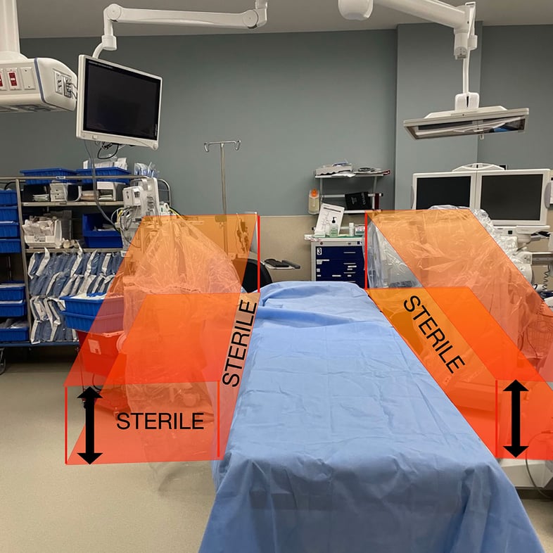
Conventional techniques try to prevent contamination of the surgical field by covering the C-arm with a disposable drape sheet that hangs from the operating field. This drape sheet is either replaced for each change in position of the C-arm or attempts are made to reuse the sheet by repetitively clipping it to the surgical drape. Such techniques have been shown to introduce contamination to the sterile field and are therefore incompatible with organizational recommendations for maintaining sterility (Gershkovich 2016, AORN 2006).
When using a new drape sheet for each C-arm position change it is not uncommon to go through 10 or more sheets, which comes at a significant supply cost. Additionally, it is my experience that often there are not enough drape sheets present in the operating room, thus making it necessary for the circulating nurse to exit the OR to go find additional sheets in the sterile supply room. This activity not only results in delays but increases foot traffic and air turbulence in the OR, which are risk factors for surgical site infections (Alizo 2019).
These considerations stand in addition to my concerns regarding compromise in the integrity of the surgical drape caused by repetitive clamping of the hanging covering sheet and formation of micro-perforations in the surgical drape. Traditional C-arm draping techniques also result in vents being formed between the down-drape and the surgical drape due to an imperfect seal that allows air turbulence to push contamination into the surgical field as the C-arm is rotated to a horizontal position.
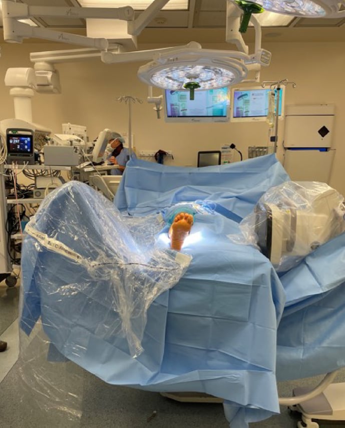
The C-Armor Drape obviates these challenges with a direct adhesive seal to the surgical drape and can be deployed with hook-and-loop fasteners to completely cover the X-ray tube circumferentially. I use one C-Armor Drape per case, no matter how many times the C-arm position is changed.
Saving Time with the C-Armor Drape
In addition to reducing surgical site contamination, the C-Armor Drape’s ability to drive OR efficiency is powerful. This benefit is clearly illustrated when considering the surgical management of tibia fractures using an intramedullary nail, a technique that relies upon the use of intraoperative fluoroscopy. During these cases, repetitive repositioning of the C-arm in the horizontal plane is required to ensure maintenance of fracture reduction and appropriate hardware positioning.
The total fluoroscopy time for surgical management of tibial shaft fractures is variable but typically ranges from 72 to 130 seconds (Williamson 2018, Kirousis 2009). Each image requires 1 to 2 seconds of fluoroscopy time, so it is not unusual for more than 50 images to be obtained over the course of a case. When I perform tibial shaft fracture cases, the total length of time I use fluoroscopy is on par with that reported. Typically, I make 15 C-arm position changes during these cases.
Given the number of C-arm position changes, maximizing the efficiency of each change can result in significant time savings. In this regard, the C-Armor Drape has proven effective in my practice. I have found that the C-Armor Drape is 8 seconds faster to deploy and retract than a reusable clip drape (i.e., 3 seconds vs. 11 seconds). Furthermore, I routinely deploy and retract the C-Armor Drape by myself whereas the traditional clip-drape technique requires two people.
Over the course of a typical tibia intramedullary nail surgery, during which the C-arm is rotated into the horizontal position 15 times, I deploy and retract the C-Armor Drape 30 times in total. In these cases, the C-arm often needs to be moved along the length of the bed to image the area of interest (e.g., knee or ankle). With a half-drape, additional repositioning is typically required to keep the C-arm covered. Thus, the C-Armor Drape saves me 2 to 5 minutes compared to traditional C-arm draping methods.
The aforementioned time savings realized with the C-Armor Drape are applicable to other surgeries I commonly perform, including ankle fracture surgery and ankle replacement surgery. These surgeries have similar C-arm position change requirements with average total fluoroscopy times of 45 and 77 seconds, respectively (Salvia 2015, Angthong 2014).
Some studies indicate 1 minute of operating room time costs upwards of $100, although the literature notes that reported values vary widely (Childers 2018). Ultimately, every minute of OR time is valuable to my practice. The time I save by using the C-Armor Drape adds up quickly and is substantial.
Final Thoughts
The C-Armor Drape’s ability to optimize sterility adherence in the operating room was the reason I first chose to use it. The markedly improved workflow efficiency this device provides in terms of time savings and ergonomic design compared to traditional draping techniques has been proven in my practice. The C-Armor Drape excels in the economic landscape of today’s value-driven healthcare system.
The C-Armor Drape is one of the most impactful devices that I have incorporated into my practice. It should be a staple in any orthopaedic surgeon’s operating room where fluoroscopy is used.
TIDI Products encourages you to request a sample of the C-Armor Drape for your ambulatory surgery center or hospital today.
BIBLIOGRAPHY
Alizo G, Onayemi A, Sciarretta JD, Davis JM. Operating Room Foot Traffic: A Risk Factor for Surgical Site Infections. Surg Infect (Larchmt). 2019 Feb/Mar;20(2):146-150. doi: 10.1089/sur.2018.248. Epub 2019 Jan 16. PMID: 30648925.
Angthong C, Adams SB, Easley ME, DeOrio JK, Nunley JA 2nd. Radiation exposure in total ankle replacement. Foot Ankle Int. 2014 Nov;35(11):1131-6. doi: 10.1177/1071100714548062. Epub 2014 Aug 26. PMID: 25160822.
AORN Recommended Practices Committee. Recommended practices for maintaining a sterile field. AORN J. 2006 Feb;83(2):402-4, 407-10, 413-6. doi: 10.1016/s0001-2092(06)60171-3. PMID: 16544860.
Childers CP, Maggard-Gibbons M. Understanding Costs of Care in the Operating Room. JAMA Surg. 2018 Apr 18;153(4):e176233. doi: 10.1001/jamasurg.2017.6233. Epub 2018 Apr 18. PMID: 29490366; PMCID: PMC5875376.
Gershkovich GE, Tiedeken NC, Hampton D, Budacki R, Samuel SP, Saing M. A Comparison of Three C-Arm Draping Techniques to Minimize Contamination of the Surgical Field. J Orthop Trauma. 2016 Oct;30(10):e351-6. doi: 10.1097/BOT.0000000000000619. PMID: 27124823.
Kaska SC. A standardized and safe method of sterile field maintenance during intra-operative horizontal plane fluoroscopy. Patient Saf Surg. 2010 Dec 13;4(1):20. doi: 10.1186/1754-9493-4-20. PMID: 21144027; PMCID: PMC3016287.
Kirousis G, Delis H, Megas P, Lambiris E, Panayiotakis G. Dosimetry during intramedullary nailing of the tibia. Acta Orthop. 2009 Oct;80(5):568-72. doi: 10.3109/17453670903350057. PMID: 19916691; PMCID: PMC2823322.
Peters PG, Laughlin RT, Markert RJ, Nelles DB, Randall KL, Prayson MJ. Timing of C-arm drape contamination. Surg Infect (Larchmt). 2012 Apr;13(2):110-3. doi: 10.1089/sur.2011.054. Epub 2012 Mar 22. PMID: 22439783.
Salvia JC, de Moraes PR, Ammar TY, Schwartsmann CR. FLUOROSCOPY DURATION IN ORTHOPEDIC SURGERY. Rev Bras Ortop. 2015 Dec 6;46(2):136-8. doi: 10.1016/S2255-4971(15)30228-7. PMID: 27027000; PMCID: PMC4799182.
Teisberg E, Wallace S, O'Hara S. Defining and Implementing Value-Based Health Care: A Strategic Framework. Acad Med. 2020 May;95(5):682-685. doi: 10.1097/ACM.0000000000003122. PMID: 31833857; PMCID: PMC7185050.
Whitehouse JD, Friedman ND, Kirkland KB, Richardson WJ, Sexton DJ. The impact of surgical-site infections following orthopedic surgery at a community hospital and a university hospital: adverse quality of life, excess length of stay, and extra cost. Infect Control Hosp Epidemiol. 2002 Apr;23(4):183-9. doi: 10.1086/502033. PMID: 12002232.
Williamson M, Iliopoulos E, Williams R, Trompeter A. Intra-operative fluoroscopy time and radiation dose during suprapatellar tibial nailing versus infrapatellar tibial nailing. Injury. 2018 Oct;49(10):1891-1894. doi: 10.1016/j.injury.2018.07.004. Epub 2018 Jul 5. PMID: 30017180.